Engineers are available to assist.

The future depends on rapidly detecting harmful chemicals in counterfeit drugs.
The opioid and counterfeit drug crisis is not slowing down. Unfortunately, overdoses from illegal drugs are hard to detect until it is too late. Spectroscopy technology using diamond-turned mirrors, beamsplitters, and other optical components is especially helpful for the detection of harmful chemical compounds that may be found in counterfeit opioids and other pharmaceutical drugs.

Spectroscopy is the study of the interaction between matter and electromagnetic radiation. In some spectroscopy applications, light emission and absorption from radiation are used to detect certain molecular compounds in samples.
There are several different types of spectroscopy including Raman, Fourier-transform infrared spectroscopy (FTIR), and infrared (IR) absorption. Raman spectroscopy is based on the scattering of light from the rotational or vibrational transition of molecules in a sample.
So few photons experience the Raman effect that acceptable spectra are produced only with long exposures or with very intense light sources like lasers.1 The wavelengths emitted are the same as the incident light, except for a very small percentage. This small amount of radiation is collected by a lens and passed through a collimator, then an entrance slit, which limits the incoming polychromatic light. A mirror then collimates the light onto a grating or other dispersing element. The grating can also help filter out the laser wavelength. Another mirror focuses the selected wavelength range onto the detector (Figure 1).

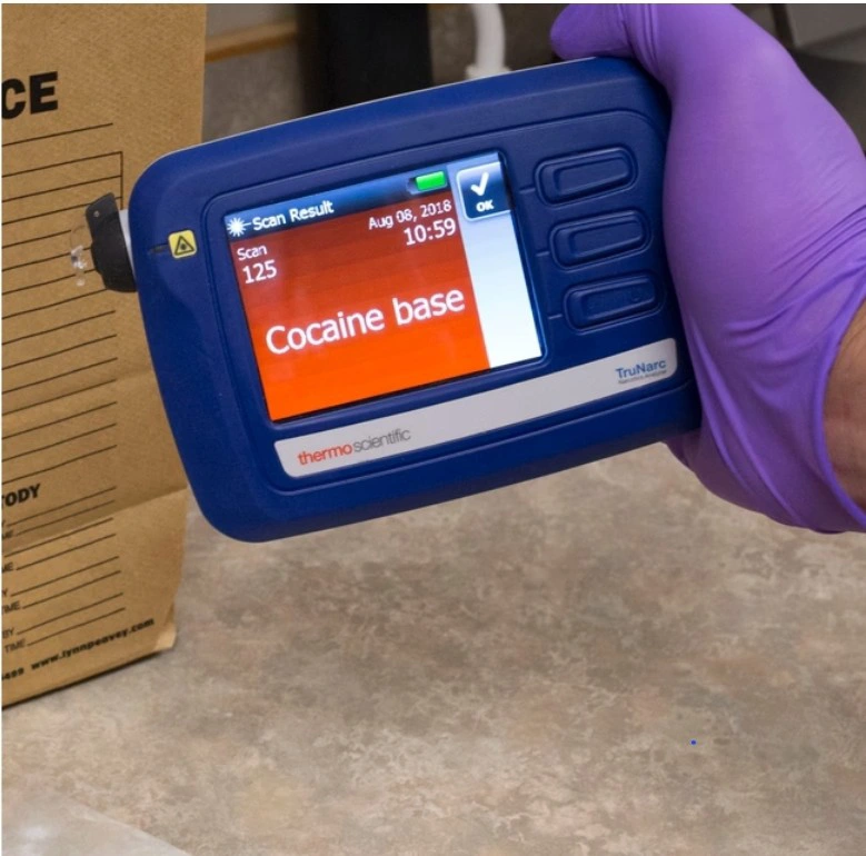
Forensic scientists use spectroscopic techniques to locate certain chemical compounds often found in counterfeit street drugs. Raman devices are advantageous for field use because of their speed, cost efficiency, and non-destructive analysis.2 Handheld versions can be carried into the field and used to quickly identify common isomers (Figure 2). Isomers are molecules with the same molecular formula, but different spatial arrangements of the atoms.3 In illicit drugs, isomers are also referred to as drug analogs, as they are used to imitate a certain drug. These analogs are often undetectable until recognized by spectrometers. Certain isomers, such as fentanyl, carfentanil, and butyrfentanyl, are found in illicit narcotics and can range from being half as potent to 10,000 times as potent as their non-analog counterparts.2
Although Raman spectrometers are fast and effective for onsite testing, they cannot catch every compound, and samples should be brought into a lab for further testing using an FTIR spectrometer.
FTIR is a subcategory of spectroscopy used in several life science applications. The name is derived from mathematician and physicist Jean Baptiste Joseph Fourier, who developed the method of transforming a function into an alternate form consisting of cosine and sine functions of different frequencies. It is commonly used in FTIR analysis.
FTIR spectrometers are utilized to distinguish chemical compounds found within substances. The main components of these machines are a source, interferometer, sample compartment, detector, amplifier, A/D convertor, and computer.4 The setup of the components can be seen in Figure 3 below.

The interferometer used is typically of a Michelson interferometer configuration consisting of a broadband light source, two mirrors, and a beamsplitter. It splits one initial beam of light in half, sends one beam towards a fixed mirror, sends the other towards a moving mirror, and then recombines them. The recombined beam passes through the drug sample to a detector. By adjusting the position of the moving mirror, individual wavelengths can be periodically transmitted or blocked because of destructive interference due to the optical path difference of the two beams.4 This allows the spectrometer to change the spectrum of the source to see what wavelengths are absorbed by the sample. A Fourier transform uses the recorded transmission for each mirror position to determine the absorption per wavelength, which identifies the chemical makeup of the sample.
In the lab, scientists can use the precise beam-splitting technology FTIR Spectrometers provide to decipher between certain isomers and deuterated analogues of drugs. The spectrometer outputs the background detected wavelengths in an interferogram, then collects a single-beam spectrum of the sample. This contains absorption bands from the sample as well as the background. These two graphs are analyzed and are finally merged into one graph as a ratio of a single-beam sample spectrum and background spectrum to show the complete sample spectrum (Figure 4).4

Not only can the FTIR spectrometers detect harmful drug compounds, they play a crucial role within the pharmaceutical industry. Pharmaceutical companies are required to follow strict testing and guidelines for production throughout product development and manufacturing.5

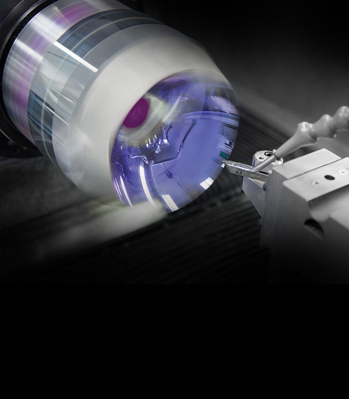
Edmund Optics provides a variety of optical components for FTIR spectrometer systems including diamond turned off-axis parabolic mirrors and beamsplitters. Our diamond turning facility is led by experts with 10+ years of experience diamond turning a wide variety of materials including metals, crystalline materials, and plastics. Edmund Optics also manufactures high-precision beamsplitters for both prototyping and volume production. FTIR spectrometers, along with countless other optics-enabled technologies, are helping create a safer and healthier future.


or view regional numbers
QUOTE TOOL
enter stock numbers to begin
Copyright 2023 | Edmund Optics, Ltd Unit 1, Opus Avenue, Nether Poppleton, York, YO26 6BL, UK
California Consumer Privacy Acts (CCPA): Do Not Sell or Share My Personal Information
California Transparency in Supply Chains Act
This content may include material that has been generated or modified using artificial intelligence (AI).
The FUTURE Depends On Optics®